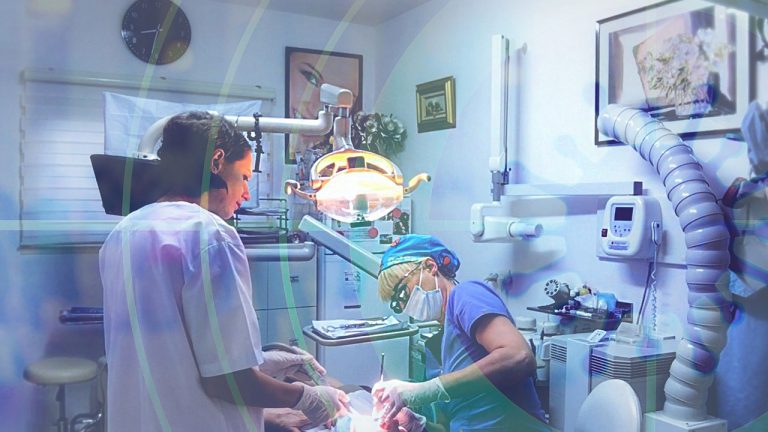

Radiology involves the studying of the human body from images generated by radiographic devices. Imaging technology like x rays have been around since the late 1800s. Xrays generate images by quickly exposing the body to a fast moving stream of electrons that are stopped by a metal plate and are developed into an image. Use of xray accessories like lead glasses and lead aprons is important for the safety of patients and practitioners.
Radiographers also perform other types of imaging studies including fluoroscopy, tomography, mammography, ultrasounds, nuclear medicine and magnetic resonance imaging. Not all of these imaging studies involve the use of radiation, but those that do should mean use of radiation protection products like lead glasses. After these images are developed a Radiologist may study the resulting images on pacs monitors for diagnostic purposes. A radiologist is a doctor who has received additional training in imaging procedures and interpolation for diagnostic purposes.
Xrays are not only used to diagnose broken bones, museums have put the technology to work to see beyond the surfaces of a painting. Wearing lead glasses, imaging studies show when artists have reused canvases and show the preliminary steps taken to achieve final masterpieces. Xrays of famous works like the Mona Lisa show there was least one other version under the surface of the painting now known around the world . This is a great source for more.


